Cloudy Swelling Kidney Pathology
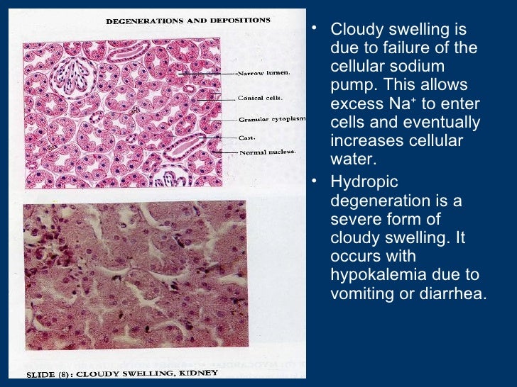
The morphological change called cloudy swelling is actually hydropic or vacuolar degeneration. We can see cloudy swelling the tubular cells are swelled and no longer has the normal morphology.
Kmu Pathology Lab Slide 1 Cloudy Swelling Kidney

Sometimes vacuoles are not present in the affected cells.

Cloudy swelling kidney pathology. 1996 Johns Hopkins School of Medicine. The cytoplasm of affected hepatocyte appears diluted or rarefied due to the presence of excessive amount of water. Brief Descriptions H ydropic change or vacuolar degereration.
Insufficiency of the supply of nutriment. Cellular swelling synonyms. Parenchymal degeneration in kidney Staining.
In advanced stages the organ appears as though it had been boiled. Image of epithelial edema cloudy - 121506972. I n 1852 and 1858 cloudy swelling was described by Virchow 1 as a condition in which a parenchymatous organ such as the liver or the kidney is swollen more cloudy and of a more doughy consistency.
This change has been termed ballooning degeneration or cloudy swelling Figure 412. Picture of Cloudy swelling of the renal tubules in kidney light micrograph photo under microscope stock photo images and stock photography. Instead the cytoplasm is diluted and organelles are widely dispersed within the rarefied electron-lucent cytoplasm.
Liver hepatitis hypoxia kidney shock myocardium hypoxia phosphates. Parenchymatous Degeneration Or Cloudy Swelling 1. NoteNote normal tubulesnormal tubules 4.
GROSS FINDINGS Affected organ Kidney Liver or heart enlarged due to swelling Cut surface- bulges outwards and is slightly opaque MICROSCOPIC FINDINGS 1. Photo about Cloudy swelling of the renal tubules in kidney light micrograph photo under microscope. B There may be increased.
The failure of the cell to make use of the material placed at its disposal is probably the more important cause. Appears whenever cells are incapable of maintaining ionic and fluid homeostasis. Thyroid hormones acute iron overload and the pathogenesis of cloudy swelling.
A The blood-supply may be actually diminished. Electron micrograph of a renal tubular epithelial cellElectron micrograph of a renal tubular epithelial cell 6. Symptoms may include sudden or intense pain in the back or side vomiting painful urination blood in the urine weakness and fever due to a urinary.
It is an intracytoplasmic accumulation of water due to incapacity of the cells to maintain the ionic and fluid homeostasis. Prominent pathological changes observed in liver were severe vascular and sinusoidal congestion with diffuse degenerative changes and mononuclear infiltration into peripheral areas while the kidney showed vascular and glomerular congestion cloudy swelling of tubular epithelium. Normal kidneyNormal kidney.
Cloudy swelling Kidney. Narrowing of hepatic sinusoids due to the swelling of hepatocyte. Hydropic change vacuolar degeneration cellular edema is an acute reversible change resulting as a response to nonlethal injuries.
Hydronephrosis is a condition of the urinary tract where one or both kidneys swell. It is easy to be observed in parenchymal organs. Gross Findings P allor increased turgor and increased in weight.
The first manifestation of almost all forms of sell injury. Cell swelling cytoplasm contains coarse granules microvasculature compressed 2Small clear vacuoles seen in cellsVACUOLAR DEGENERTION. Anyone can become affected by.
This happens because urine does not fully empty from the body. Acute cellular oedemaAcute cellular oedema cloudy swelling Hydropic degeneration cloudy swelling Hydropic degeneration 5. Hydronephrosis swollen kidney is a result of urine build-up within one or both kidneys making them swell and enlarged.
شرائح عملي الباثولوجي طب الزقازيق 2018 Basiounitis. 1Institute of General Pathology and CNR Centre for Research in Cell Pathology University of Milan Italy. Marked cell swelling of renal tubular epithelial cells.
This severe cellular edema is classically called hydropsroot word meaning water hence hydropic change.

Cloudy Swelling Of The Renal Tubules In Kidney Light Micrograph Stock Photo Picture And Royalty Free Image Image 105112485

Mykoweb Toxic Fungi Of Western North America

F Kidney Histopathological Examination Of Den Tu Group Higher Download Scientific Diagram
General Pathology Lecture 1 Introduction Cell Injury

Cloudy Swelling Of The Renal Tubules In Kidney Stock Photo Image Of Epithelial Edema 121506972
Kmu Pathology Lab Slide 1 Cloudy Swelling Kidney

Cloudy Swelling Of The Liver Light Micrograph Stock Photo Image Of Inflammation Magnification 157930532
Kmu Pathology Lab Slide 1 Cloudy Swelling Kidney


Gp 4 Liver Of Mice Showing Severe Congestion Arrow Cloudy Download Scientific Diagram

Cloudy Swelling Of The Liver Light Micrograph Stock Photo Image Of Pathology Edema 157930122

Cloudy Swelling Of The Liver Light Micrograph Stock Image Image Of Injury Micro 157931143

Gnps Treated Rat Received 100 Ml Of 10 Nm Particles For 7 Days Download Scientific Diagram

Cell Swelling An Overview Sciencedirect Topics

Cloudy Swelling Renal Image Photo Free Trial Bigstock
Photomicrograph Of The Kidney Showing A B H E Renal Corpuscles Download Scientific Diagram
Kmu Pathology Lab Slide 1 Cloudy Swelling Kidney
Kmu Pathology Lab Slide 1 Cloudy Swelling Kidney

Cloudy Swelling Of The Liver Light Micrograph Stock Image Image Of Infection Cloudy 157930861
Http Www Bu Edu Eg Portal Uploads Veterinary 20medicine Pathology 2392 Crs 11496 Files Mohamed 20mahmoud 20salem 20gaballah 20 Cell 20swelling Pdf
Post a Comment for "Cloudy Swelling Kidney Pathology"