Atrophic Kidney Pathology Outlines
Sustained Decreases in urine output oliguria salt and water overload rising BUN concentrations hyperkalemia and other manifestations of uremia. Hard GC Alden CL Bruner RH Frith CH Lewis RM Owen RA Krieg K Durchfeld-Meyer B.
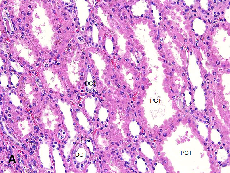
Kidney Tubules And Collecting Ducts American Urological Association
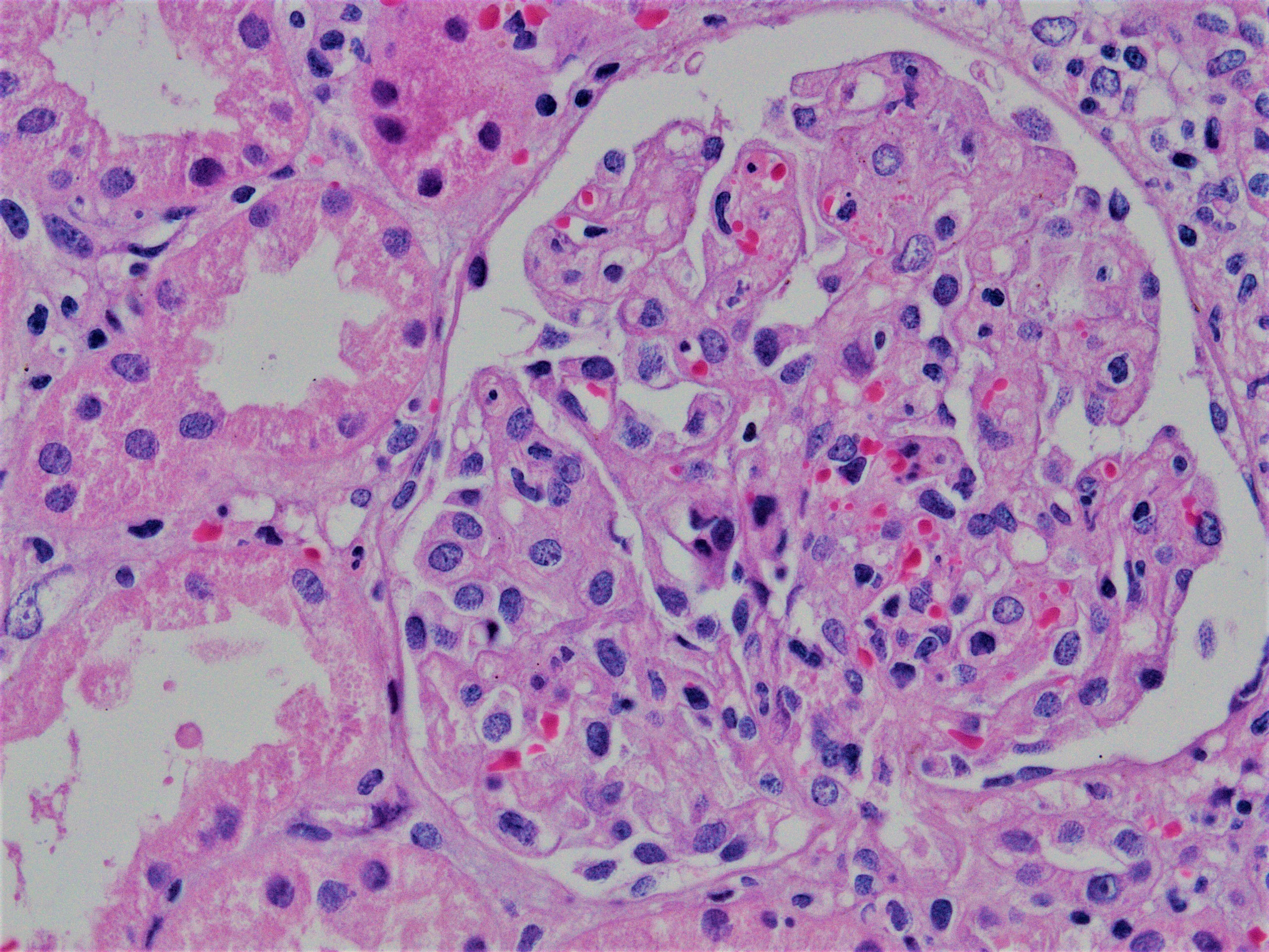
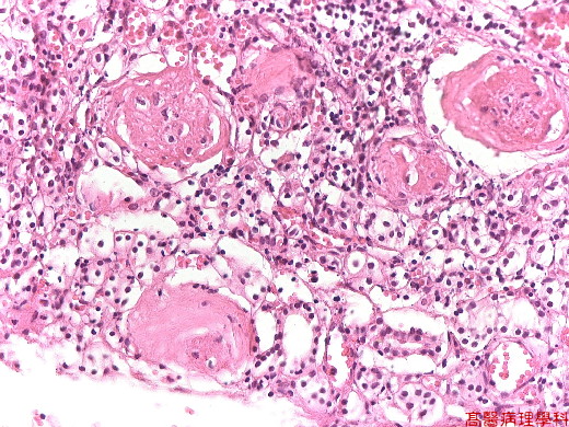
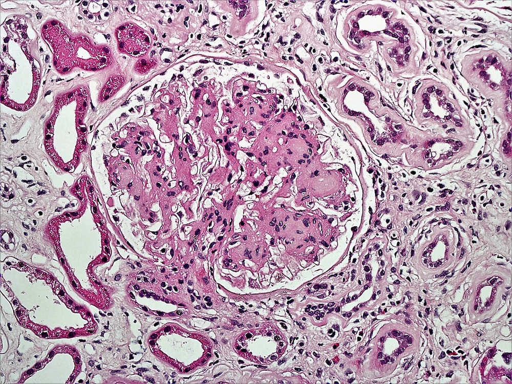
Advanced segmental sclerotic lesions are present in these two glomeruli with increased mesangial matrix and obliteration of capillaries and with more extensive lesion in the glomerulus on the right.

Atrophic kidney pathology outlines. Increased extent of tubular atrophy and accompanying interstitial fibrosis correlates with worse prognosis. An atrophic kidney is one that has shrunk to an abnormal size with abnormal function. Obstructive uropathy is structural or functional hindrance of normal urine flow sometimes leading to renal dysfunction obstructive nephropathy Acute obstruction increases susceptibility to infection.
Peter CP Burek JD Van Zwieten MJ. Cysts dilated tubuli and colector ducts lined by cuboidal or flattened epithelium contain an eosinophilic fluid. We include in this review the following new and emergingprovisional renal entities.
Spontaneous nephropathies in rats. We include in this review the following new and emergingprovisional renal entities. Between the cysts the intervening parenchyma is reduced atrophic by compression.
- Initial polyuria as water balance is restored. This is also known as renal atrophy. Dilation is characterized by distention and dilation of the renal pelvisusually accompanied by renal papilla atrophy Figure 1 and Figure 2.
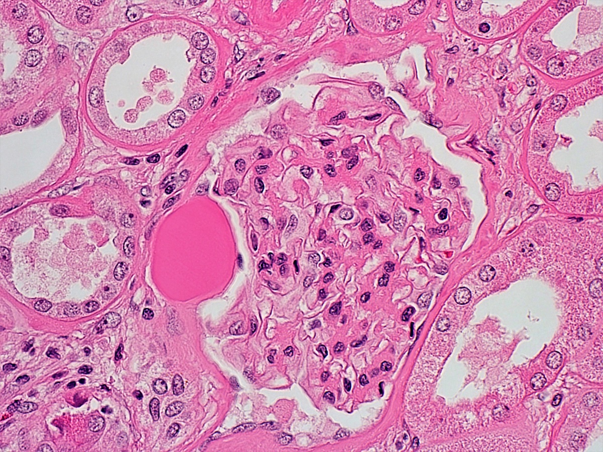
The lesions are typical of idiopathic FSGS. WebPathology is a free educational resource with 11226 high quality pathology images of benign and malignant neoplasms and related entities. What is the recovery phase of ATI.
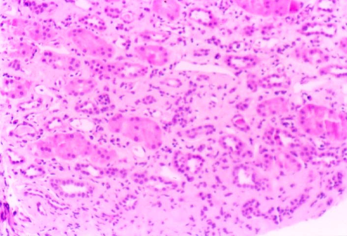
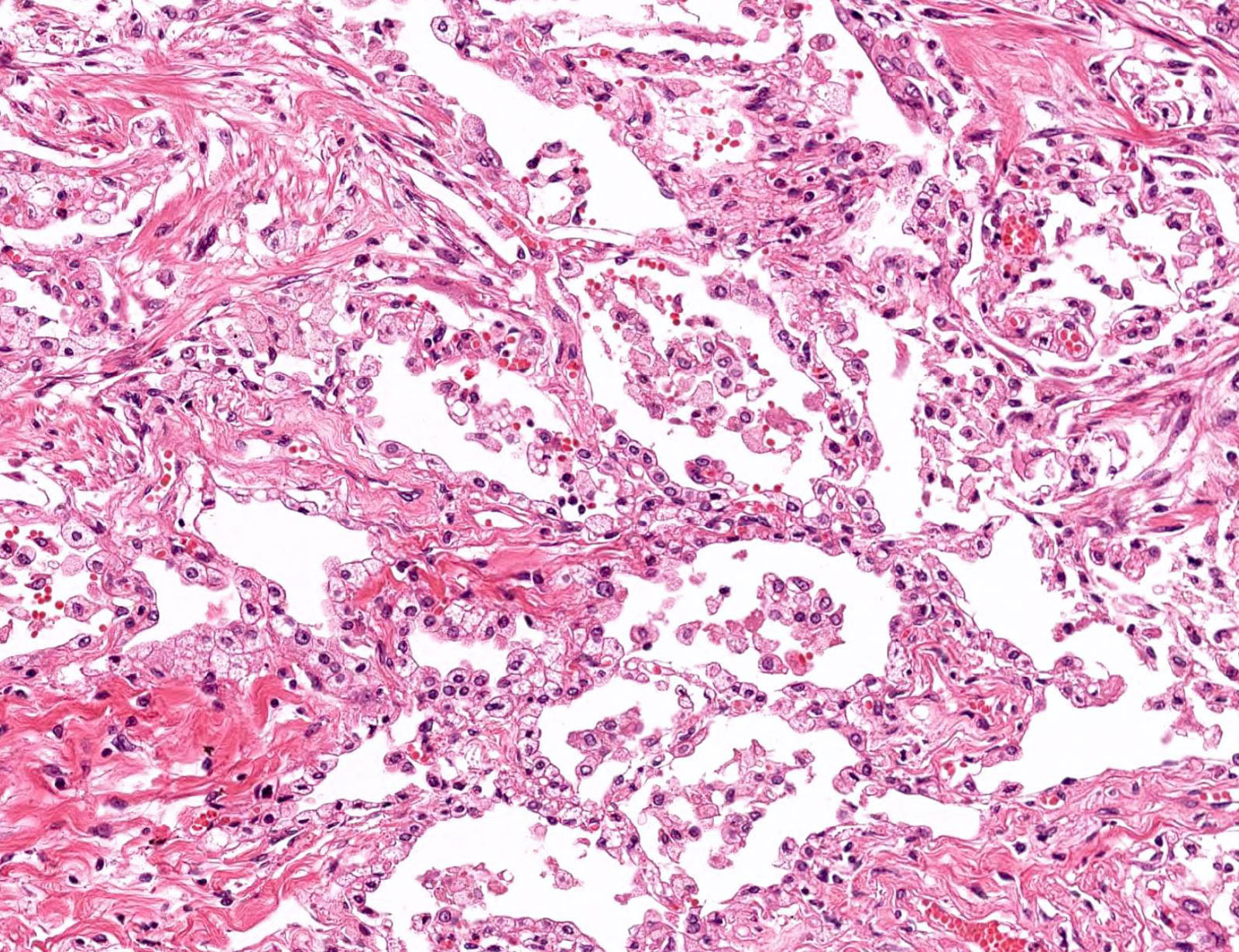
Non-proliferative lesions of the kidney and lower urinary tract in rats. Page views in 2021 to date this page and chapter topics. One of the many patterns of tubular atrophy in the kidney is the aptly named thyroidization pattern because of its resemblance to normal thyroid gland follicles.
STPARPAFIP Washington DC 1-32. Succinate dehydrogenase-deficient renal cell carcinoma thyroid-like follicular carcinoma of the kidney anaplastic lymphoma kinase rearrangement-associated renal cell carcinoma renal cell carcinomas with prominent smooth muscle stroma fumarate hydratase-deficient renal cell carcinoma biphasic squamoid papillary renal cell carcinoma eosinophilic solid and cystic renal cell carcinoma atrophic kidney. Tomsich Institute of Pathology and Laboratory Medicine.
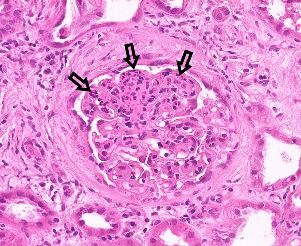
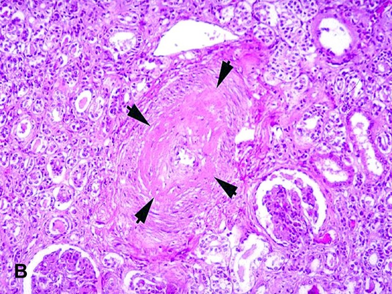
In summary we describe the clinicopathologic features of 8 unique benign atrophic kidney-like lesions that may simply represent a non-neoplastic form of organizing tubular atrophy and glomerulocystic change and emphasize their distinction from thyroid-like follicular carcinoma of the kidney. The interstitium show moderate to severe fibrosis and tubular atrophy. Chronic obstruction causes hydronephrosis.
The atrophic cortex is characterized by reduced thickness of the one or more of the cortical layers due to a decrease in cell size or a loss of cells. Tubular atrophy is a general term that describes several patterns of chronic tubular injury with thickened tubular basement membranes and clinically manifests as chronic kidney disease with decreased glomerular filtration rate. Visual survey of surgical pathology with 11226 high-quality images of benign and malignant neoplasms related entities.
There is variably decreased overall size of the gland often with distortion of the gland outline. In summary we describe the clinicopathologic features of 8 unique benign atrophic kidneylike lesions that may simply represent a non-neoplastic form of organizing tubular atrophy and glomerulocystic change and emphasize their distinction from thyroid-like follicular carcinoma of the kidney. Cleveland Clinic Robert J.
A greater decrease in kidney size especially for both kidneys can lead to kidney failure. Guides for Toxicologic Pathology. - Renal function begins to improve rapidly with resolution in a few weeks.
The zonae fasciculata and reticularis are more often affected than the zona glomerulosa. Chronic infections or blockage of the kidney can also result in kidney atrophy. Dilation of the renal pelvis is preferred over the term hydronephrosiswhich can denote either a gross necropsy or microscopic change.
Bladder neck obstruction congenital meatal stenosis anterior posterior urethral valves Pediatr Surg Int 200925613 ureteropelvic. A kidney that is smaller in size can lead to kidney disease. The dilated tubules contain abundant protein resembling thyroid colloid which is surrounded by flattened epithelial cells.
However microscopically this parenchyma is represented by functional nephrons. This type of kidney atrophy is due to a lower blood supply to the kidney sandor loss of nephrons the basic working units of the kidneys. Succinate dehydrogenasedeficient renal cell carcinoma thyroidlike follicular carcinoma of the kidney anaplastic lymphoma kinase rearrangementassociated renal cell carcinoma renal cell carcinomas with prominent smooth muscle stroma fumarate hydratasedeficient renal cell carcinoma biphasic squamoid papillary renal cell carcinoma eosinophilic solid and cystic renal cell carcinoma atrophic.
Its not the same thing as renal.
Pathology Outlines Acute Chronic Active T Cell Mediated Rejection
Transplant Pathology Internet Services
Pathology Outlines Iga Nephropathy
Kidney Nonneoplastic Lesion Atlas

Kmu Pathology Lab Slide 129 Hydronephrosis Kidney
Pathology Outlines Oxalosis
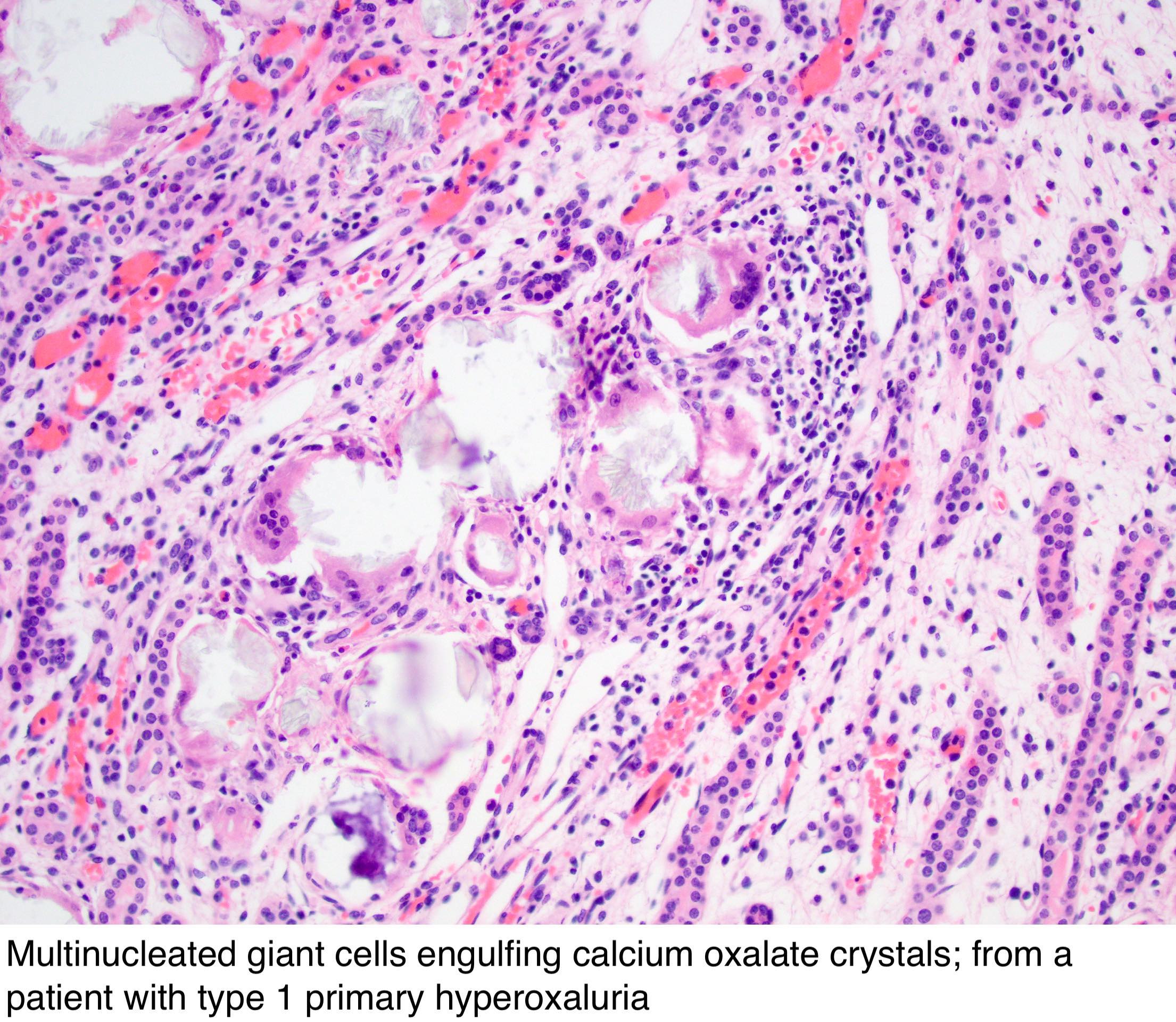
Pathology Outlines Oxalosis

Kmu Pathology Lab Slide 129 Hydronephrosis Kidney
Chapter 3 Tumours Of The Stomach Pathology Outlines

Pathology Outlines Diabetic Kidney Disease

Pathology Outlines Diabetic Kidney Disease
Pathology Outlines Diabetic Kidney Disease
Malignant Hypertension American Urological Association
Renal Pathology Renal Pathology Acute Renal Failure

Pathology Outlines Covid 19
Chronic Glomerulonephritis Medical School Studying Chronic Histology Slides

Renal Pathology

Renal Pathology Pathology Cancer Cancer Prevention
Poststreptococcal Acute Diffuse Proliferative Glomerulonephritis Hypercellular Medical Education Histology Slides Diffuser

Post a Comment for "Atrophic Kidney Pathology Outlines"