How To Read A Panoramic X Ray
A difference is that it is captured using a side-to-side sweeping motion instead of the full 360 degree non-stop motion used in panoramic X-rays. This makes it possible for your dentist to notice any of the following problems.

Introduction Panoramic Radiographs Technique Anatomy Review Continuing Education Course Dentalcare Com
A panoramic may provide lots of information about other thingsthe position of wisdom teeth for example but dont depend on it to tell you much about the joints.

How to read a panoramic x ray. Mandible Maxilla Zygoma Soft tissue Air spaces Teeth Interpreting Panoramic Radiographs Examine the borders of the bone first Next examine the medullary bone Check the internal structures such as canals foramina and sinuses. Panoramic radiography is a form of focal plane tomography. A panoramic x-ray machine has two sides.
Why cavities show up on x-rays. One containing an x-ray tube and the other containing a film or detector. Panoramic X-ray reveals cysts tumors bone irregularities and much more.
During a panoramic x-ray well position your head using chin forehead and side rests. The procedure to take a panoramic dental x-ray requires the patient to rest their chin on a platform while lightly biting into a grooved plastic piece that denotes the center of your mouth and props your mouth open slightly. Panoramic x-ray shows the dentist the patients nose area sinuses lower and upper jaw joints teeth and the surrounding bone structure.
5 oral orifice. Unlike other kinds of dental x-ray which capture just one tooth or a section of the mouth panoramic x-rays show the whole mouth in one single image. Since decay is an area of tooth demineralization an area of reduced mineral content or even possibly an outright hole a space that would have no mineral content at all those locations where it has formed will show as a darkened area on an x-ray.
Panorex ie panoramic x-ray is also very necessary in planning dental implant surgeries to be performed by dentists. B external ear. Well also stabilize your bite to keep your mouth slightly open.
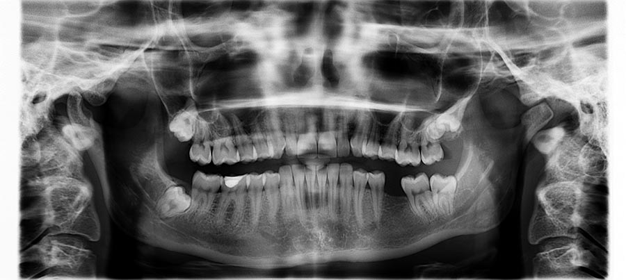
Then the x-ray machine will rotate in a semicircle around your head allowing us to get a comprehensive picture of your entire mouth and jaw. Thus images of multiple planes are taken to make up the composite panoramic image where the maxilla and mandible are in the focal trough and the structures that are superficial and deep to the trough are blurred. Why Use a Panoramic X-Ray.
The technical term for a panoramic dental x-ray is an orthopantomogram so you can see why its referred to as an OPT or OPG x-ray. The first step in understanding panoramic anatomy is to appreciate the perspective from which each part of the image is presented. Cephalometric dental x-ray Ceph A two dimensional x-ray image that captures images of the entire skull and side profiles.
Other nonproprietary names for a panoramic radiograph. A external nose. A panoramic dental X-ray creates an image of your entire mouth including the upper and lower jaws all the teeth temporomandibular TMJ joints and even your nasal area and sinuses.
See Figures 5 and 6 for details. 1 nasal fossa. 4 oral cavity.
In this introductory video we talk about how dental x-rays work how to read them and how to apply the buccal object rule to localize objects in an x-ray i. A panoramic radiograph is a panoramic scanning dental X-ray of the upper and lower jaw. Cephalometric Analysis is an X-ray similar to a panoramic X-ray in that it has the capability of capturing a full view of your skull and neck.
It shows a two-dimensional view of a half-circle from ear to ear. This is because the x-ray beam is directed from a position about 30 degrees under the joint and portions of the condyle are projected upward and superimposed over other boney structures in the image. When complete a Ceph looks like the image seen here.
Thats because the decayed portion of the tooth is less hard less dense or intact and therefore the x. These are positioned on a rotating arm which revolves 180 degrees around your head to capture the full image. Your head is supported while the arm rotates to capture the image.
Panoramic radiograph with major soft tissue structures af and airways 15 traced. Interpreting Panoramic Radiographs Examination is an orderly process. Because the image is captured by an X-ray tube which rotates around the patients head rather than from a stationary source this perspective changes from the posterior regions of the jaws to the anterior area.
D lingual tonsils on posterior tongue. F soft palate.
Optimal Panorex Imaging Decisions In Dentistry
Optimal Panorex Imaging Decisions In Dentistry
Panorex Whats A Panorex Why Do I Need That Dr Nima Massoomi Dmd Med Md
12 Tomography And Panoramic Radiography Pocket Dentistry

Panoramic Radiograph Landmarks Tutorial Part 2 Youtube

Panoramic Radiograph An Overview Sciencedirect Topics
Https Onlinelibrary Wiley Com Doi Pdf 10 1111 J 1834 7819 2011 01655 X
Https Onlinelibrary Wiley Com Doi Pdf 10 1111 J 1834 7819 2011 01655 X
Can A Panorex Dental X Ray Detect Tmj Problems


Interpretation Of Panoramic Radiographs Perschbacher 2012 Australian Dental Journal Wiley Online Library

Interpretation Of Panoramic Radiography With Interactive Cases Youtube
Panorex Whats A Panorex Why Do I Need That Dr Nima Massoomi Dmd Med Md
What Are Digital X Rays Are They Safer Than Film X Rays
Optimal Panorex Imaging Decisions In Dentistry

Analysis Of A Panoramic Radiograph University Of Toronto Faculty Of Dentistry Information And Instructional Technology Services

Bc Oral Pathology Case No 48 Extra Extra Read All About It Dentistryiq

Interpretation Of Panoramic Radiographs Perschbacher 2012 Australian Dental Journal Wiley Online Library

Technology My Family Dentist
Https Onlinelibrary Wiley Com Doi Pdf 10 1111 J 1834 7819 2011 01655 X
Post a Comment for "How To Read A Panoramic X Ray"